


Application
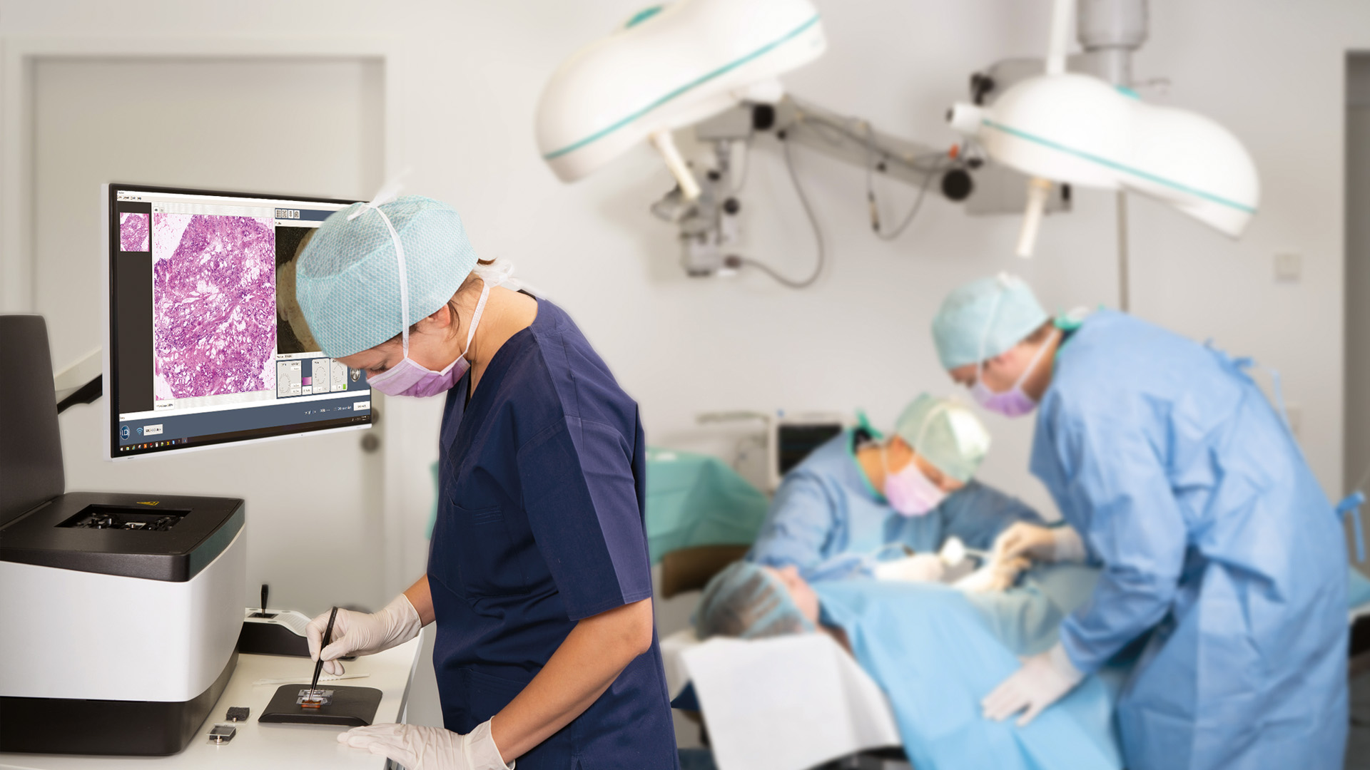
The VivaScope 2500 is suitable for various types of applications. For example intraoperative assessment of tumor margins and immediate examination of biopsies. Surgical workflows and patient management can thus be significantly improved. The acquired images show subcellular details of the examined tissue and provide information similar to H&E staining.
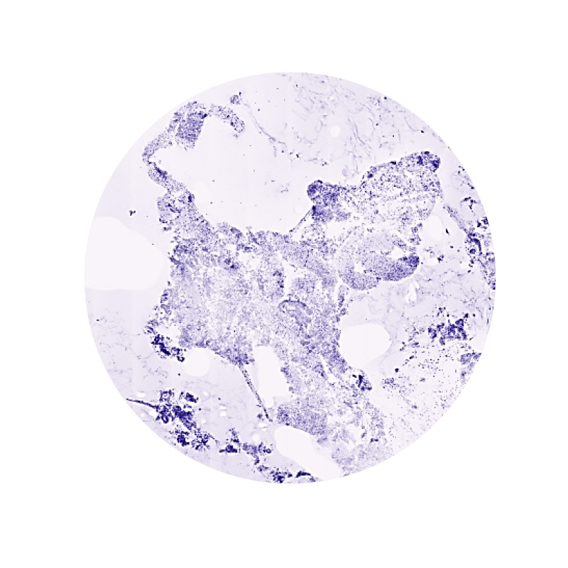
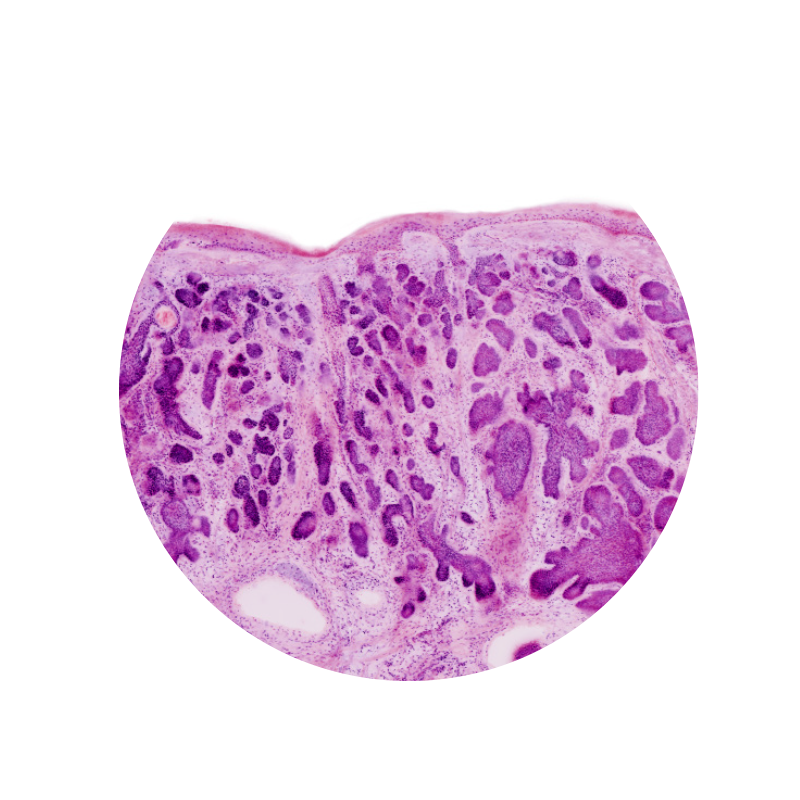
1. FNA / FNB and small tissue fragments
CytoMatrix is a novel, patented technology for the collection and preservation of FNA/FNB samples and small tissue fragments. In combination with the VivaScope 2500, it revolutionizes the handling and analysis of cytological and microhistological specimens. The analysation and adequacy assessment of these samples can be rapidly performed while maintaining the integrity of the specimen for subsequent histological, immunohistochemical and molecular analysis.
That means for you:
✔ Minimal preparation
✔ Full tissue preservation
✔ Optimized ressource allocation
✔ Efficient patient managment
✔ Advanced patient care
✔ Remote evaluation
Image courtesy of Dr. Anna Crescenzi, University Hospital Campus Bio-Medico, Rome
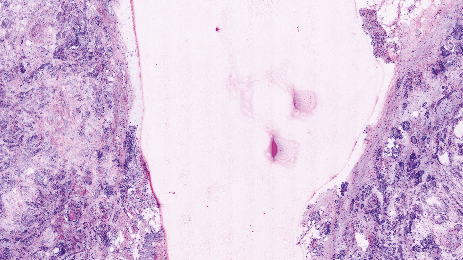
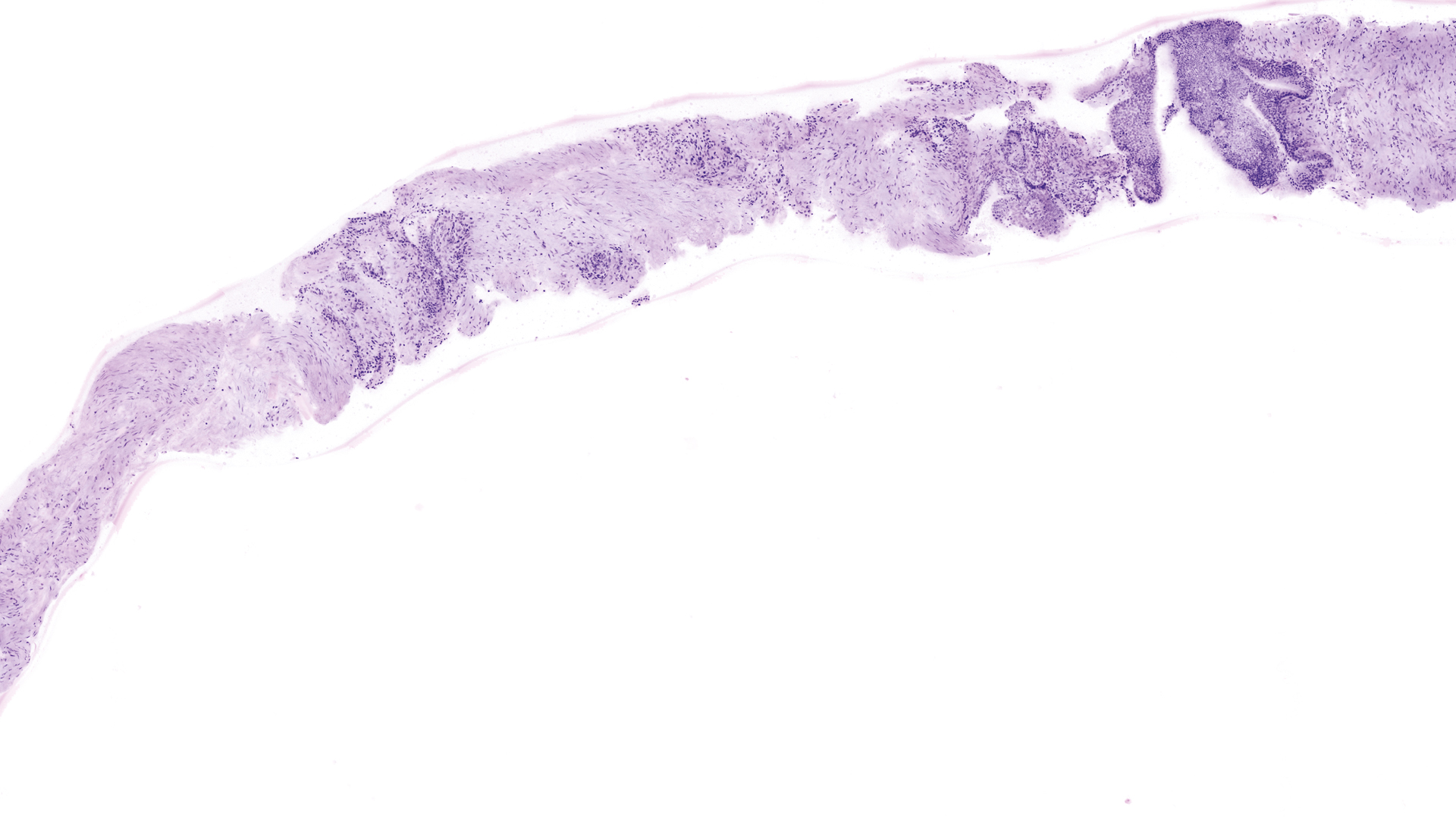
2. Intraoperative margin control
The VivaScope 2500 technology offers many advantages over frozen section analysis for microscopically controlled surgery. The time needed to complete a surgery can be reduced significantly. Integrated into a surgical workflow, VivaScope scans provide information comparable to H&E images derived from FFPE or frozen sections. The examination can be performed without a laboratory.
That means for you:
✔ No laboratory required
✔ Remote evaluation
✔ Advanced patient care
✔ Improved patient turnaround time
Image courtesy of Dr. Anna Crescenzi, University Hospital Campus Bio-Medico, Rome
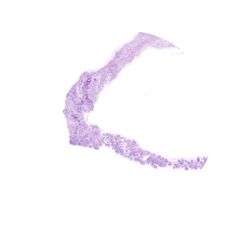
3. Core biopsies
The processing and imaging of core biopsies takes less than 5 minutes and the results can be evaluated instantly. The conclusions drawn from the examination can have a direct impact on the patient’s treatment, e.g. enabling therapy to be scheduled immediately, thus within a single hospital stay.
That means for you:
✔ Rapid evaluation at the bedside
✔ Optimize biopsy acquisition
✔ Reduce biopsies or avoid re-biopsies
✔ Immediately initiate treatment schedule
Image courtesy of Dr. Stefano Puliatti, Dr. Laura Bertoni, Dr. Paola Azzoni, University of Modena and Reggio Emilia
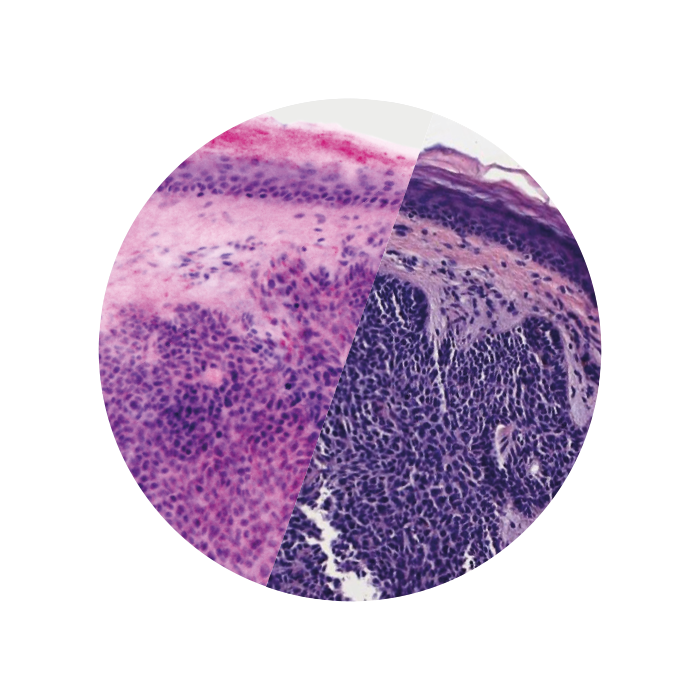
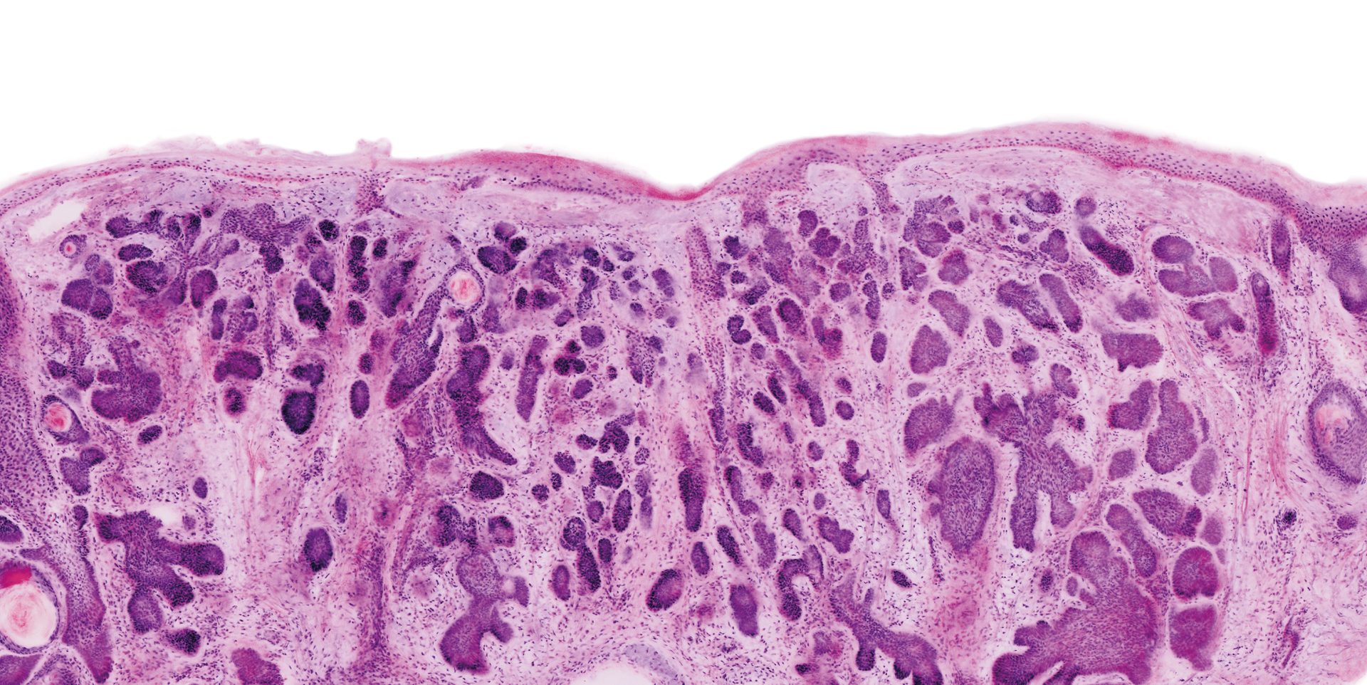
H&E-like images. At the bedside during surgery. Within 5 minutes.
Two lasers of different wavelengths create two distinct images, a fluorescence image and a reflectance image. Both signals are scanned simultaneously and are used to create pseudo-colored images. The device’s software uses an algorithm to translate the acquired image information into colors that resemble H&E.
Images courtesy of Dr Javiera Pérez-Anker.
Basal cell carcinoma; imaged with the VivaScope 2500 (left) and after H&E staining (right)