


Instant Digital Pathology
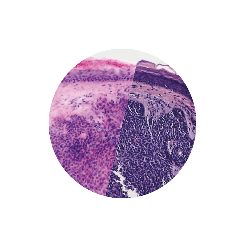
VivaScope ex vivo technology allows for direct pathological assesment during surgery. Like H&E staining, VivaScope images are generated from two components. Two lasers of different wavelengths create two distinct images, a fluorescence image and a reflectance image. Both signals are scanned simultaneously and are used to create pseudo-colored images. The device’s software uses an algorithm to translate the acquired image information into colors that resemble H&E.
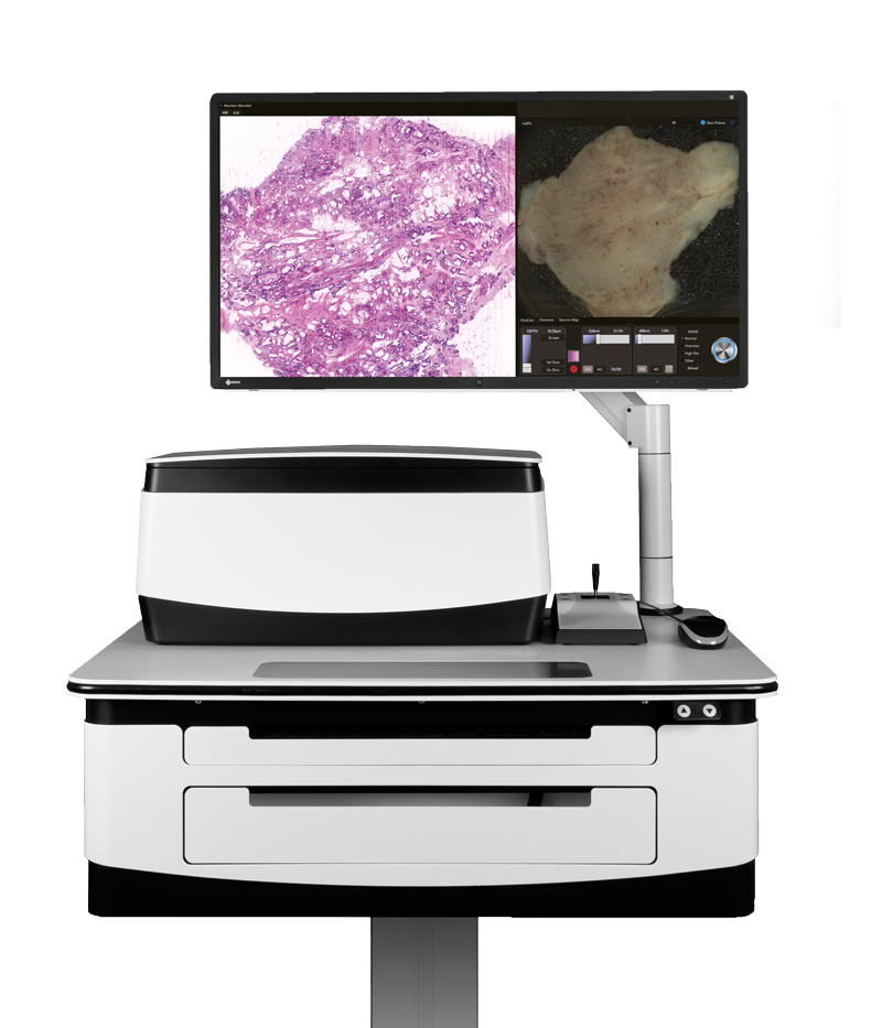
H&E-like digital images. At the bedside. Intraoperatively. In 5 minutes only.
Fresh tissue can be examined immediately after an excision without lengthy procedures. This allows for the direct assessment of the specimen in the operating room. All packed in our “moveable” laboratory, the VivaScope 2500. The device is capable of working within a hospital’s DICOM environment to enable storage, search, viewing, scheduling and backup of acquired images.
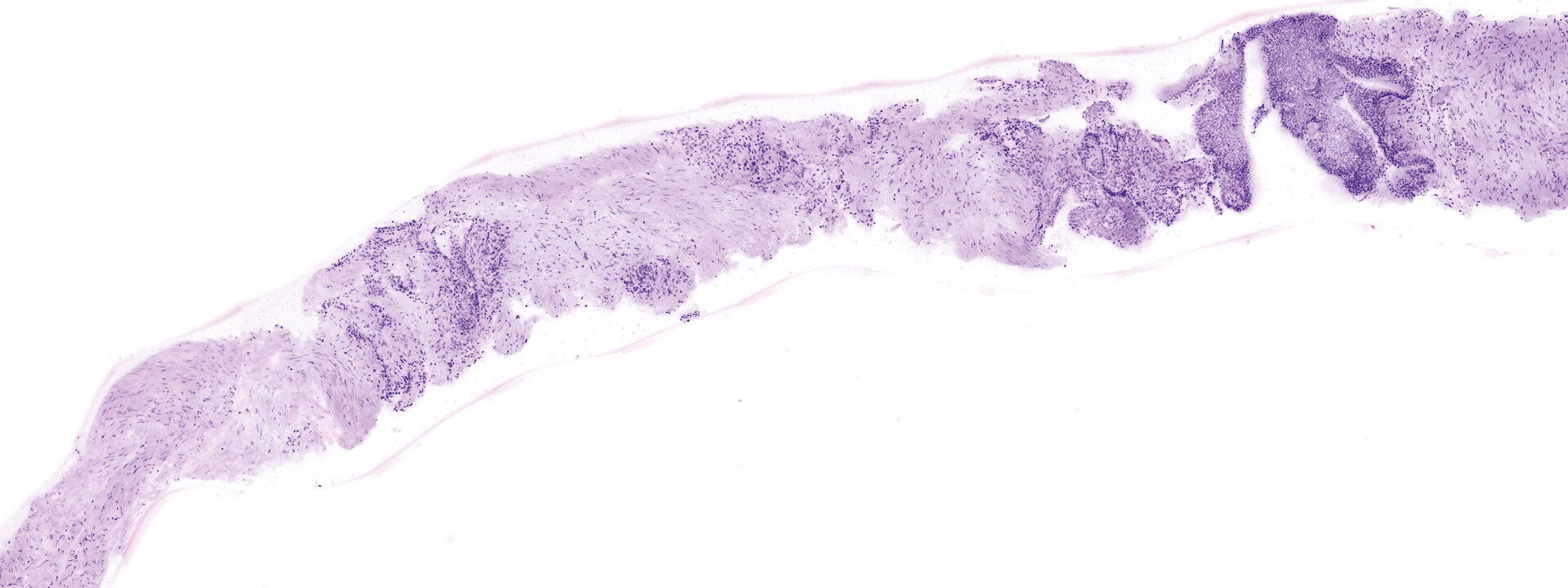
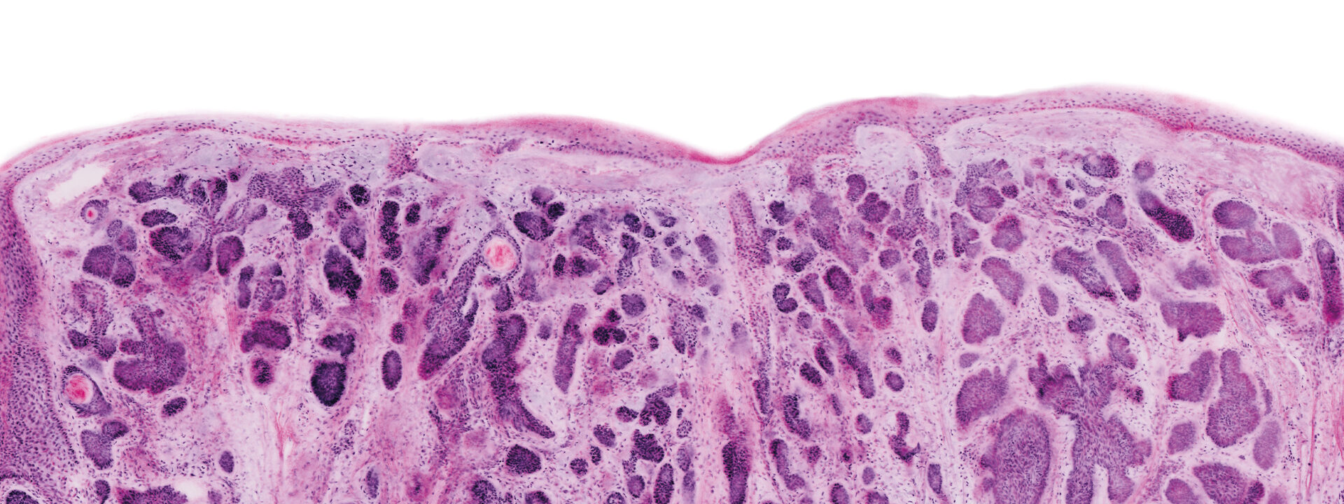
Images courtesy of Dr Javiera Pérez-Anker. Basal cell carcinoma; imaged with the VivaScope 2500 (left) and after H&E staining (right).
See every detail. Seamless zoom up to550x. With a great sample size.
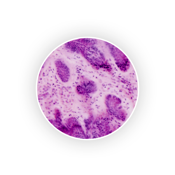
The VivaScope technology is based on confocal microscopy (CLSM) and acquires images of superb optical resolution and contrast. The VivaScope 2500 images allow seamless zoom with up to 500x magnification and a great sample size.
The microscope can be installed on a movable table and thus be used in different locations and surgery rooms in clinics / hospitals.
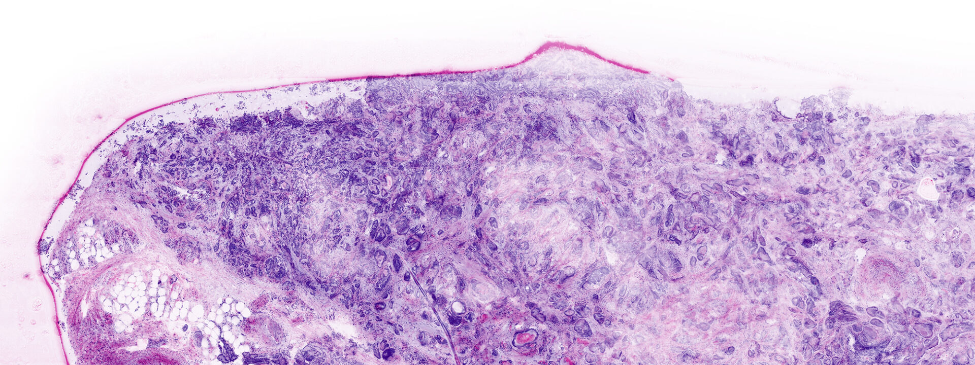
Image courtesy of Dr Javiera Pérez-Anker, Hospital Clinic of Barcelona.