


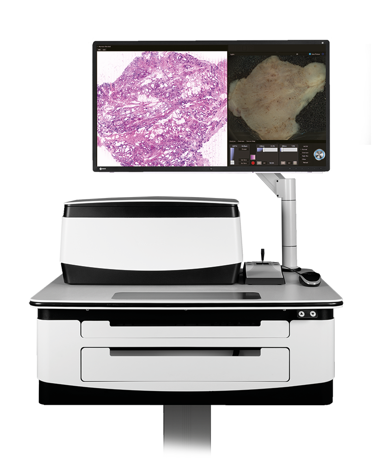
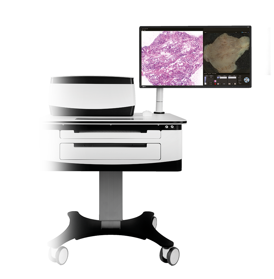
The VivaScope 2500
Your laboratory on a cart – with the VivaScope 2500 ex vivo microscope. Ready for your 5-minute workflow?

Minimal preparation & direct assessment
Remote evaluation / telemedicine
Significant time saving
Tissue integrity
About:
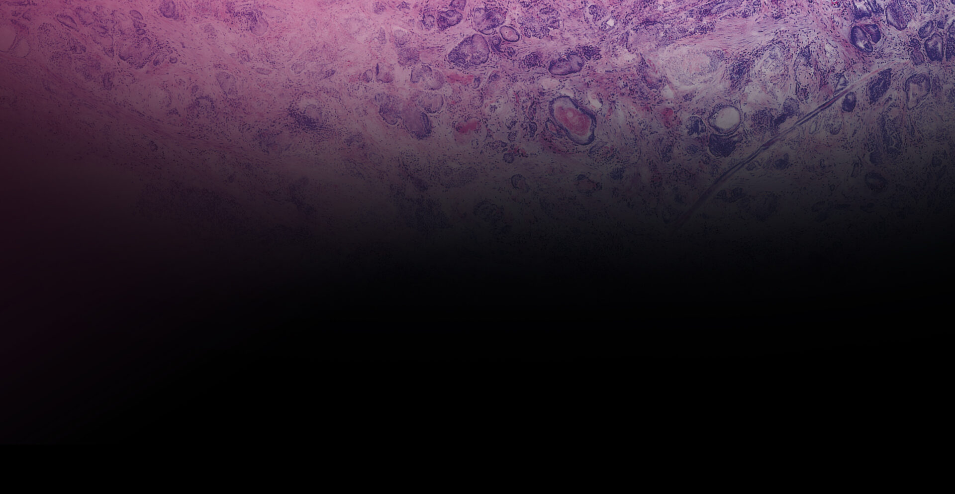
The VivaScope 2500 ex vivo technology offers H&E-like images, generated from two components. Two lasers of different wavelengths create two distinct images, a fluorescence image and a reflectance image. Both signals are scanned simultaneously and are used to create pseudo-colored images. The device’s software uses an algorithm to translate the acquired image information into colors that resemble H&E.
Access your sample morphology.. Within 5 minutes.
The VivaScope 2500 rapidly scans the excised tissue and reveals the cellular morphology right at the bedside. The examined tissue remains unharmed by the procedure and can be preserved for later histopathological analysis.

H&E-like digital images. At the bedside during surgery.
High resolution images of unfixed tissue without sectioning. Within 5 minutes, digital and on the bedside. The device’s software uses an algorithm to translate the acquired image information into colors that resemble H&E.
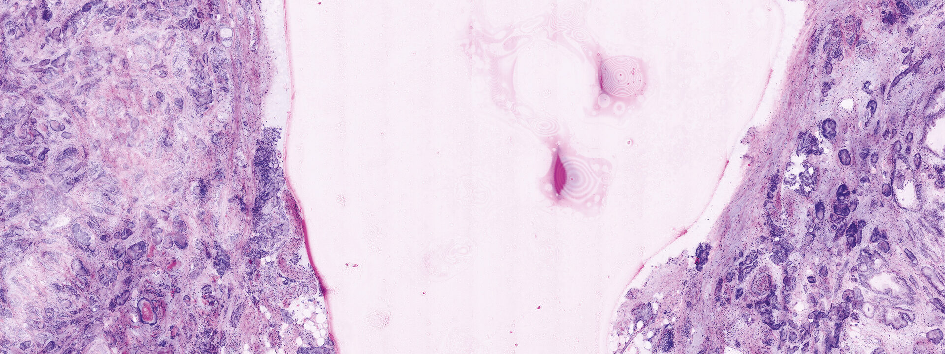
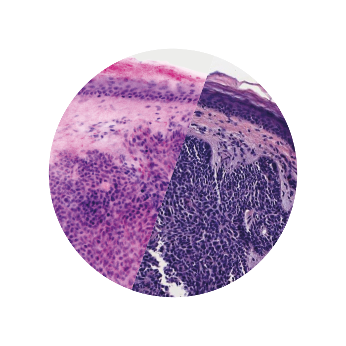
Images courtesy of Dr Javiera Pérez-Anker.
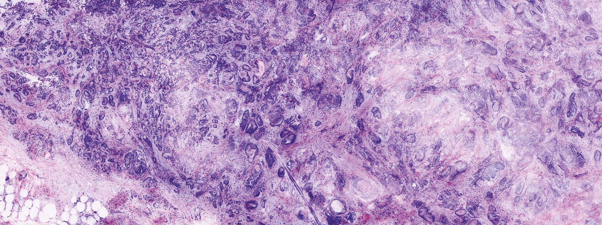
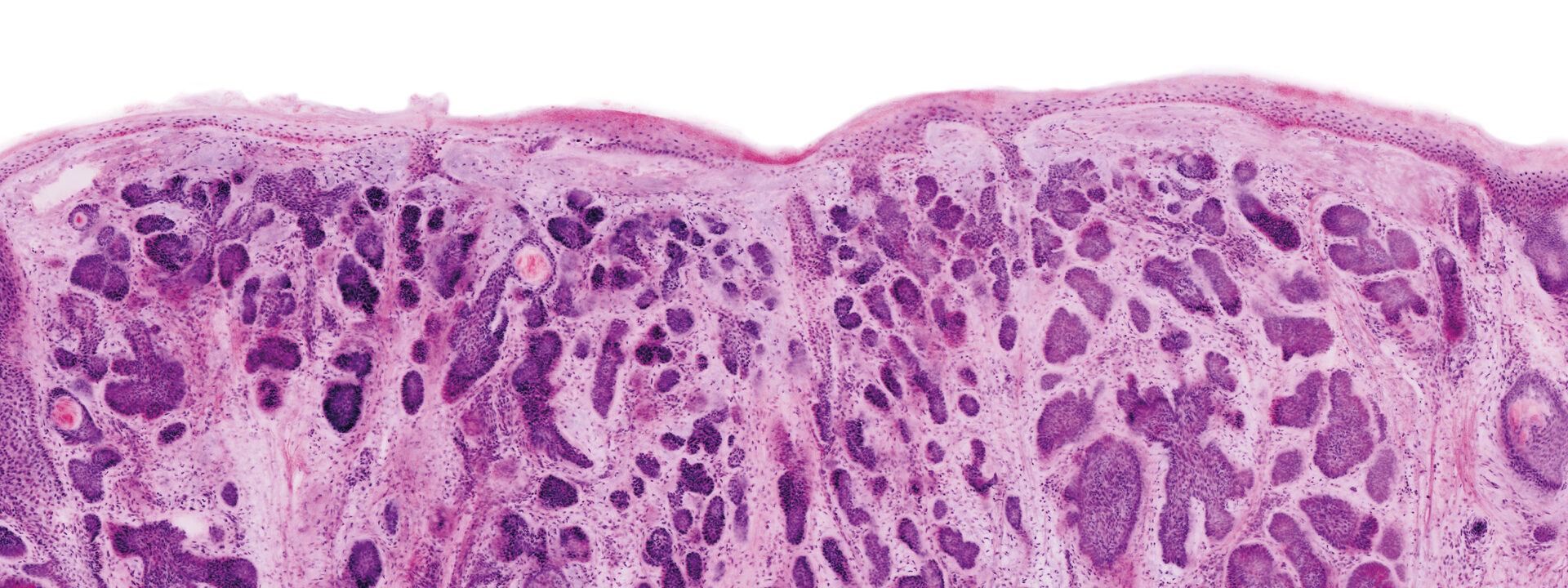
Basal cell carcinoma; imaged with the VivaScope 2500 (left) and after H&E staining (right)
Full Preservation.
The VivaScope 2500 revolutionizes the evaluation of cytological and microhistological specimens. The analysation and adequacy assessment of these samples can be rapidly performed while maintaining the integrity of the specimen for subsequent histological, immunohistochemical and molecular analysis.
Evaluation of tumor margins
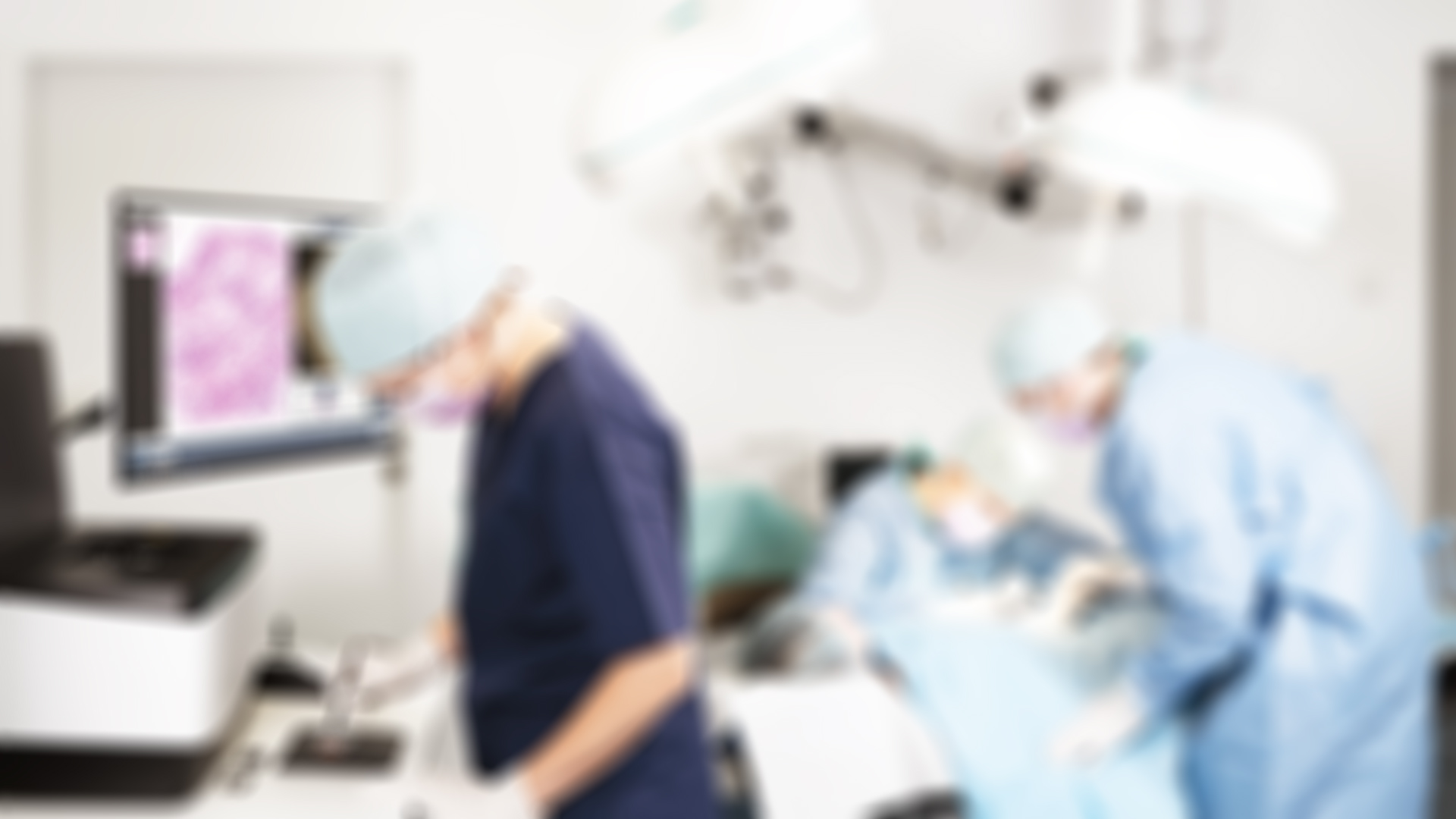
The VivaScope 2500 allows you to assess tumor margins intraoperatively. The ex vivo technology offers many advantages over frozen section analysis for microscopically controlled surgery. The time needed to complete a surgery can be reduced significantly. Integrated into a surgical workflow, VivaScope scans provide information comparable to H&E images derived from FFPE or frozen sections.
Your benefits:
✔ No laboratory required
✔ Remote evaluation
✔ Advanced patient care
✔ Improved patient turnaround time
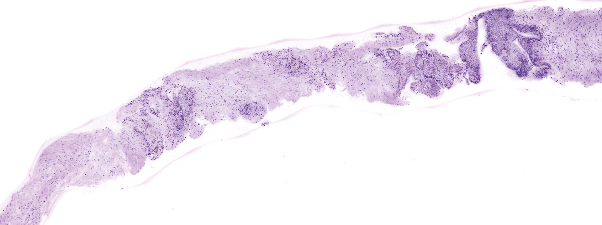
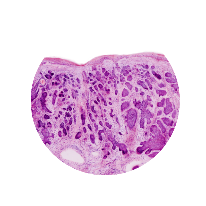
Image courtesy of Dr. Anna Crescenzi, Department of University Hospital Campus Bio-Medico, Rome.
