


Find Centers
Instant Digital Pathology
Technology
Category
Country
Your Location
Reset
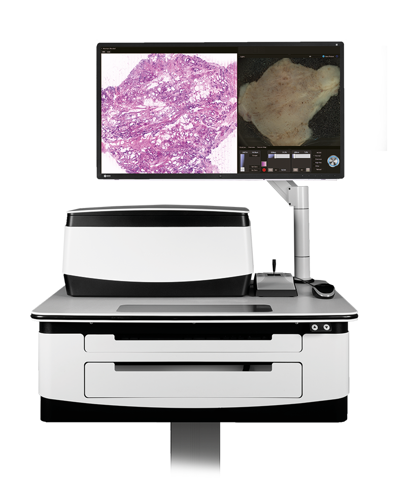
H&E-like images. At the bedside during surgery. Within 5 minutes.
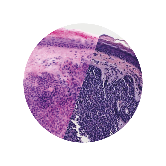
Two lasers of different wavelengths create two distinct images, a fluorescence image and a reflectance image. Both signals are scanned simultaneously and are used to create pseudo-colored images. The device’s software uses an algorithm to translate the acquired image information into colors that resemble H&E.
Images courtesy of Dr Javiera Pérez-Anker.
Basal cell carcinoma; imaged with the VivaScope 2500 (left) and after H&E staining (right)
Find Centers:
Technology
Category
Country
Your Location
Reset
Show More
Sorry, no clinic or reference centre exists (yet) for this combination.
We are constantly expanding our clinic partners.
If you have any questions, contact us.
Sorry!