


Milestone at Vienna General Hospital: VivaScope revolutionises cancer diagnosis and treatment in Austria
In Austria, around 42,000 people are diagnosed with cancer every year. Although this is a matter of health and therefore the greatest human good, the analogue frozen section – a technique first used in 1895 – is still considered the gold standard for intraoperative pathological assessment when removing tumour tissue. With its “Ex Vivo” technology, the company VivaScope is bringing about a fundamental medical revolution. Thanks to digital optical sections, tumour tissue can be removed more efficiently than ever before and assessed within five minutes using telemedicine. The VivaScope 2500 is now being used for the first time in the field of dermatology at Vienna General Hospital.
Vienna / Munich, 07.11.2023. The Austrian population is growing and ageing. This will be accompanied by an increasing number of cancer cases in the coming decades. This poses an enormous challenge for the healthcare system and medical staff. “Malignant tissue neoplasms must be confirmed by a pathologist on the basis of his preparatory, microscopic and molecular biological examinations. However, while the number of diseases will increase, the number of medical specialists for this will decrease in parallel,” explains Dr Roberto Banchi, Application Specialist and Head Of Global Application Team at the medical technology company VivaScope. This is precisely where the company comes in, using innovative technology and digitalisation to bring about a new era in the diagnosis and removal of tumour tissue.
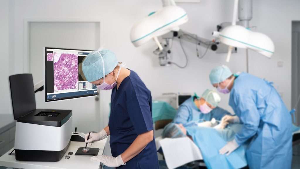
The VivaScope 2500 Ex Vivo enables the histopathological assessment of freshly removed tissue in just five minutes. “The common analogue alternatives are physical sections. With paraffin sections, however, it takes 24 hours before a result is available. Although frozen sections provide results within 20 to 45 minutes, they are less precise and produce artefacts, says Dr Banchi. With digital frozen sections, on the other hand, the tissue is stained with a fluorescent dye immediately after removal and placed on a glass plate. The tissue is then scanned with two lasers of different wavelengths, generating digital images that show the cellular morphology. The tissue remains intact thanks to the new procedure and can be used for later analysis. “The availability of meaningful results in real time saves valuable resources and protects patients from further appointments with long waiting times,” explains Dr Banchi. This is because the Ex Vivo technology immediately makes it clear whether the tissue sample taken is of sufficient quality to be analysed. If this is not the case, another sample can be taken immediately. According to the expert, the fact that the technology is also suitable for telemedical use is particularly advantageous for rural regions where a pathologist is not always present in the home.
Vienna General Hospital relies on VivaScope technology for dermatology
The digital procedure is being used for the first time in Austria at Vienna General Hospital, where a VivaScope 2500 was put into operation in mid-October. “We are already supporting the facility in the field of dermatology. The VivaScope device is sometimes used here to carry out incision margin checks during the operation,” explains the application specialist. “The critical skin area or the edge of the tumour is often removed with too large a safety radius to be on the safe side,” Dr Banchi continues. This technology is now designed to avoid this: “Because the incision margin is already checked during the operation, the doctor can act accordingly and remove more tissue if necessary. The affected area can then be closed immediately. The patient does not have to be operated on again and healthy tissue is spared,” continues the expert. These innovative microscopes from the Munich-based company VivaScope are partly produced in Austria. In addition to a production site in the USA, the company Wild in Völkermarkt (Carinthia) also manufactures components for VivaScope.
VivaScope not only has the potential to modernise the examination of removed tissue, but also manages to reduce biopsies altogether. Potentially critical skin areas can be digitally examined with the In Vivo technology, allowing even deep layers of skin to be viewed and minimising or avoiding countless biopsies. However, the technology does not only promise relief in dermatology, as Dr Banchi explains: “Numerous studies have shown that the use of VivaScope devices also brings success in other areas such as gastroenterology, urology and mammography.” VivaScope was also able to provide initial help in liver transplant pathology: the quality of donor livers is usually still analysed using frozen sectioning, which, however, results in valuable time and costs. The VivaScope Ex Vivo technology delivers results in around five minutes – if required, remote pathological evaluation is even possible in real time. As part of another large multi-centre study involving ten international centres, the VivaScope Ex Vivo technology was also able to deliver specific results for a differential diagnosis of pancreatic cancer. The company is also working on promising studies to open up further areas of application for the innovative technologies, such as lung, breast and thyroid cancer.
Motto: relieve staff, save costs
Initial figures also show that costs can be halved when using VivaScope technology, while the number of patients treated increases over the same period. On the one hand, this can reduce the number of days patients are hospitalised, which is particularly important in view of the shortage of staff in the care sector. “Secondly, some of the treatment with VivaScope devices can also be carried out by junior doctors, allowing head physicians to use their resources more efficiently,” emphasises Dr Banchi. In times of acute staff shortages in medicine and nursing, it is therefore possible to actively contribute to speeding up cancer detection processes and making the lengthy path to diagnosis as gentle as possible for those affected.
Contact person